Newsportal - Ruhr-Universität Bochum
SICM: Hochauflösende Abbildung der Oberfläche lebender Zellen
Die SICM wurde 1989 von Hasma und Kollegen entwickelt [1] und die Anwendung der SICM zur Abbildung lebender Zellen wurde zuerst 1997 von Korchev und Kollegen gezeigt [2,3]. Mit Hilfe der SICM wurden verschiedene zellphysiologische Fragestellungen untersucht, die in einem öffentlich verfügbaren Review-Artikel nachzulesen sind [4].
Funktionsweise
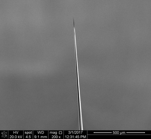
Raster-elektronenmikriskopische Aufnahme einer Glas-Nanokapillare, wie sie in der SICM zum Einsatz kommt.
SICM nutzt den Strom durch die Öffnung einer Glas-Mikro- oder -Nanokapillare (wieim Bild oben gezeigt) um den Abstand zwischen Probenoberfläche und Sondenöffnung zu bestimmen. Bei großen Abständen zwischen Probe und Sondenspitze ist der Strom nahezu konstant, und daher ist der normalisierte Strom, also der Strom normiert auf den Strom in quasi-unendlichem Abstand, gleich eins. Für kleineren Abständen nähert sich der Strom asymptotisch null an. Der genaue Verlauf der Strom-Abstands-Kurven iwird durch die geometrischen Parameter der Sondenöffnung [5] als auch durch den Winkel zwischen Sonde und Probe [6] bestimmt.
Eine Verringerung des Stroms lässt sich bereits beobachten, bevor eine Berührung der Sondenöffnung und der Probenoberfläche stattfindet. Daher erlaubt die SICM das berührungsfreie und daher passive Abtasten der Probe. In den meisten Anwendungen wird ein Schwellwert der Stromreduktion festgelegt – häufig eine Reduktion um 1% – der benutzt wird, um die Probe aufzunehmen.
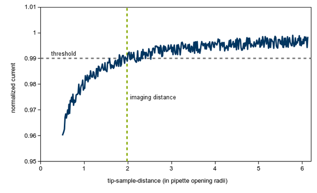
Beispielhafte Darstellung einer Strom-Abstandskurve für SICM-Aufnahmen. Ein Schwellwert von 1% (und daher ein verbleibender Strom von 99%) und der daraus resultierende Abstand, bei dem die Probe aufgenommen wird, sind als gestrichelte Linien eingezeichnet.
Beispielaufnahme
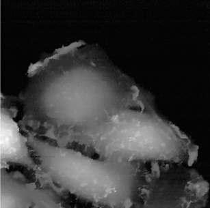
Beispielaufnahme von HeLa-Zellen. Die Abbildung ist Referenz [7] entnommen.
Copyright: The Royal Society of Chemistry. Reproduktion durch die Autorenrechte in der Publikationslizenz abgedeckt.
Literatur
- Hansma et al., Science, 1989. DOI: 10.1126/science.2464851
- Korchev et al., Journal of Microscopy, 1997. DOI: 10.1046/j.1365-2818.1997.2430801.x
- Korchev et al., Biophysical Journal 1997. DOI: 10.1016/S0006-3495(97)78100-1
- Happel et al., Sensors (Basel) 2012. DOI: 10.3390/s121114983
- Rheinlaender and Schäffer, Analytical Chemistry, 2017. DOI: 10.1021/acs.analchem.7b03871
- Thatenhorst et al., Analytical Chemistry, 2014. DOI: 10.1021/ac5024414
- Gesper et al., Nanoscale, 2017. DOI: 10.1039/C7NR04306F