
Newsportal - Ruhr-Universität Bochum
News
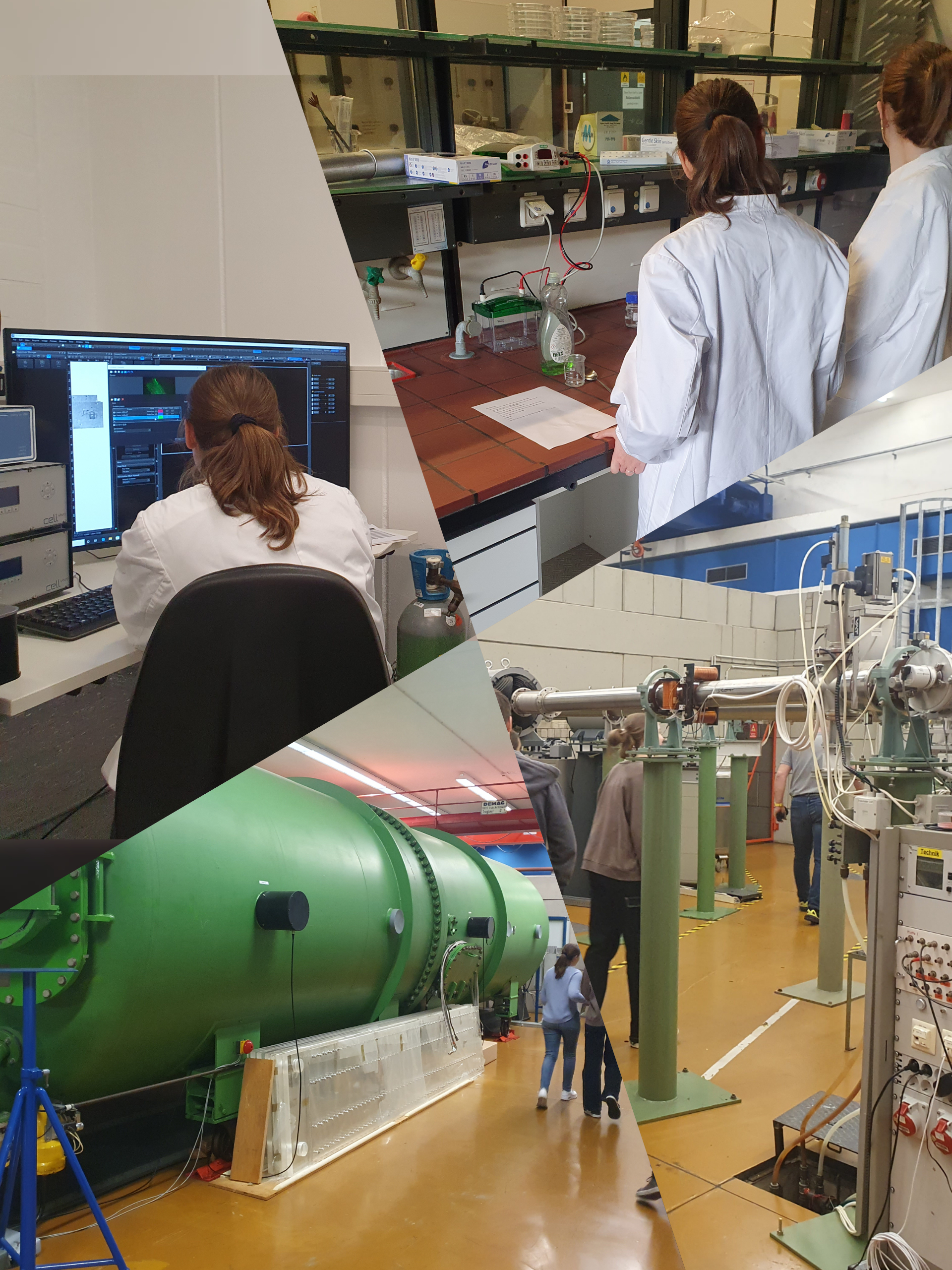
Once again, the RUBION opened its doors for this year's Girl's Day and gave two 8th grade students, Carolin and Eliane, an insight into the research taking place at RUBION.
The two got an impression of what it's like to be a scientist, were able to take a look at the particle accelerator and then put their hidden talents to the test by microscoping brain cells.
We are already looking forward to giving interested students an insight into our work again next year!
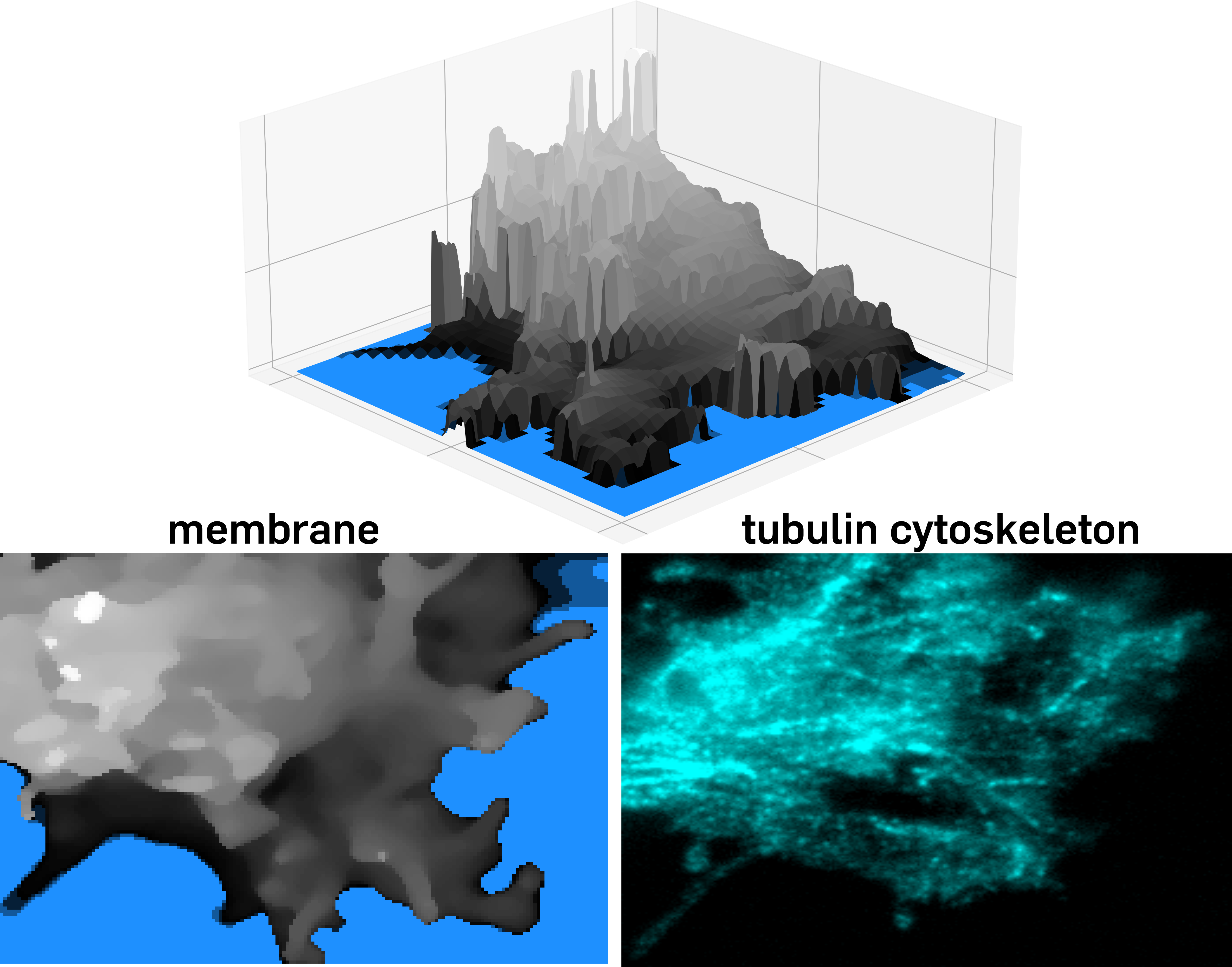
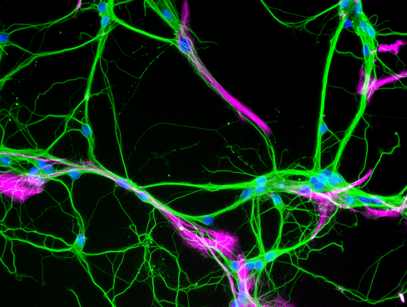
Last year, the RUBION group Nanoscopy headed by Annika Haak successfully combined expansion microscopy and scanning ion conductance microscopy. Our microscopists used this method combination to investigate the growth cones of oligodendrocyte progenitor cells. The results have been published in Biological Chemistry and are now publicly available at de Gruyter. If you have any questions about this project or are considering incorporating scanning ion conductance microscopy or expansion microscopy into one of your projects and would like to use RUBION resources, please contact rubion+nanoscopy@rub.de.
On September 14th and 19th the super-resolution microscopy facility at RUBION in cooperation with the faculty for chemistry and biochemistry participated in the Talenttage Ruhr 2023. 4 small groups of interested kids and adolescents got to learn about fluorescence microscopy and how this technique can be applied to visualize brain cells. Participants had the opportunity to capture their own micrographs of living brain cells. Furthermore, we showed the participants how 3D printed parts can be used to build a low-cost smartphone microscope.
Are you also interested to learn more about microscopy at RUBION? Send an email with your inquiry to rubion+nanoscopy@rub.de.
Form September 21st to 23rd, the RUBION group Nanoscopy presented their research at the annual meeting of the German Physiological Society in Berlin. Heiko Leßlich and Annika Haak discussed the most recent results of their projects focussing on ‘Thyroid Hormone-Induced Expression of Na+/K+-ATPase in Neural Cells Depends on Na+ Influx’ and ‘10x Cryo-Expansion Microscopy of Glial Cells’ during the meeting’s poster sessions and met great interest. The presented posters can now be found in the RUBION’s foyer on NT/NI 05.
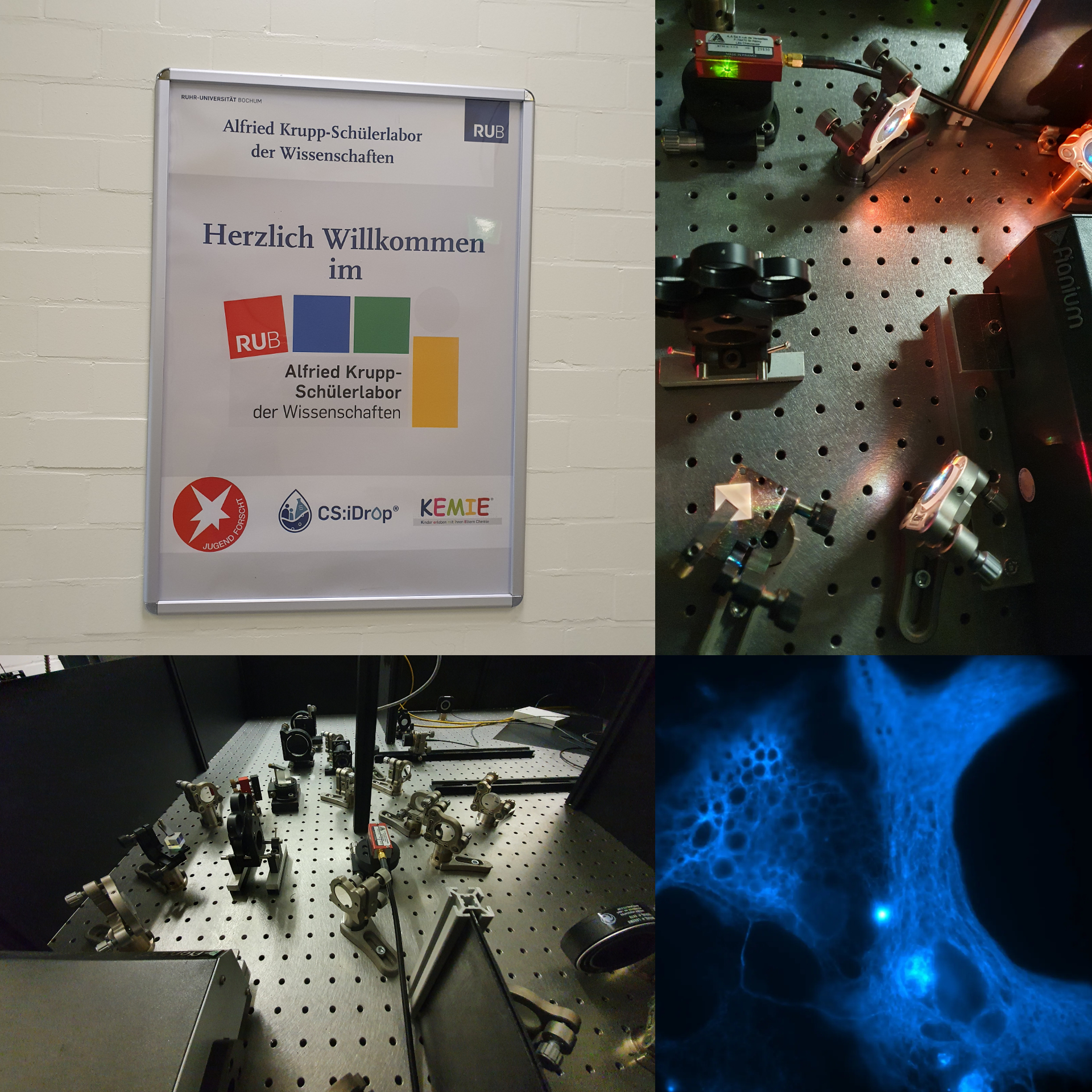
On tuesday we had the opportunity to show 13 high school students, who participates in the student project week organized by the Faculty of Physics and Astronomy, around our microscopy labs. Our nanoscopy team showed the students how STED and SIC microscopes are constructed and what can be investigated with these microscopes. Among other things, we discussed resolution limits, cell skeletons, and fluorophores. We had a lot of fun giving an insight into our work and are already looking forward to next time!
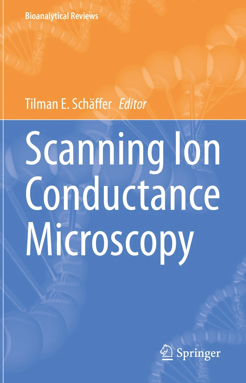
The RUBION nanoscopy research group works on, among other things, correlated STED and ion scanning conductance microscopy. Their thoughts and expertise on the setup and application of such correlated microscopes can now be found in form of a chapter in the newly published Springer Analytical Reviewś book 'Scanning Ion Conductance Microscopy' (Editor: Tilman Schäffer): Link
In cooperation with the working group Electrobiochemistry of Neural Cells (Prof. Irmgard Dietzel-Meyer, Department of Biochemistry II) the working group Nanoscopy under the leadership of Heiko Leßlich has investigated methods for the optimization of primary cell cultures. Primary cell cultures from the rat brain represent an important model for basic research on nerve cells. Since a variety of different cell types exist in the brain, cell-cell interactions can complicate research. By adding substances that inhibit proliferation of certain cell types, cultures can be enriched with neurons. The effect of two cytostatic drugs on cell culture composition was evaluated using automated cell counting of micrographs. All study details and results can be found in the open access PLOS ONE publication. For further information contact Heiko Leßlich (heiko.lesslich@rub.de).